
Bone stress injuries often begin as microscopic damage well before they show up on standard clinical X-rays or routine scans. By the time a traditional diagnosis finally happens, the injury has usually progressed enough to require weeks or months of debilitating downtime. A team of biomedical engineers hopes to change that clinical reality entirely through the application of advanced high-frequency ultrasound.
The core breakthrough relies on how sound waves travel through and bounce off trabecular bone structure. Healthy bone tissue reflects ultrasonic frequencies in a predictable, uniform pattern. When micro-cracks and early structural fatigue start forming under repetitive mechanical loading, that sonic signature alters in distinct ways. The diagnostic hardware captures these subtle shifts instantly, mapping out areas of localized bone weakness long before localized pain forces a patient to stop training.
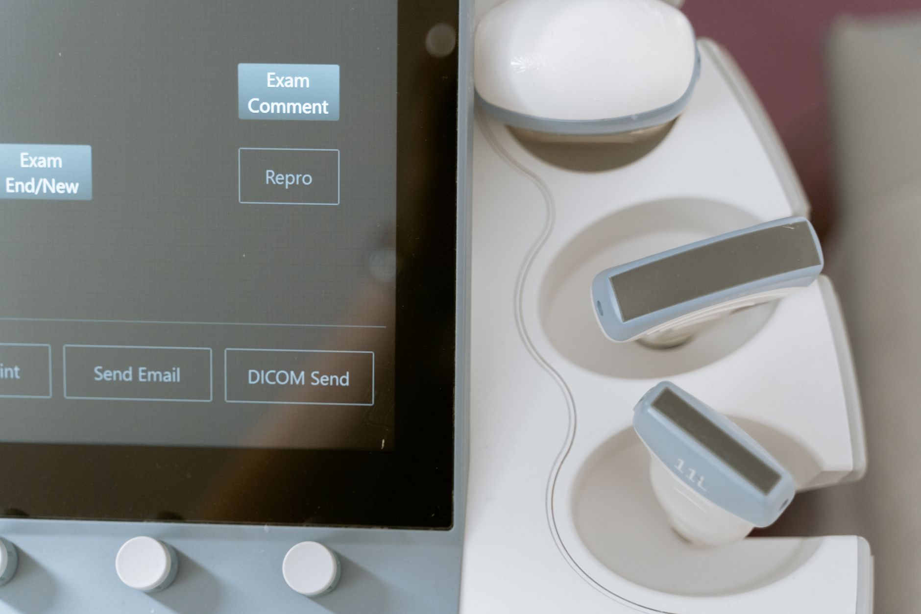
Clinical trials involving elite athletes and military recruits are already underway to validate the portability and accuracy of the device. Because the hardware can be scaled down into handheld probes, point-of-care screening could soon happen directly on sports sidelines or active-duty bases rather than inside specialized hospital radiology departments.
Medical device manufacturers are taking notice of the commercial potential, signaling that orthopedic care is moving toward proactive monitoring. Catching stress injuries at day one rather than day thirty means personalized training adjustments can prevent full fractures entirely, altering recovery timelines across professional sports and physical rehabilitation clinics worldwide.
New Ultrasonic Tech Spots Stress Fractures Long Before X-Rays Can





Comments